Minimally Invasive Image-Guided Procedures
Many conditions that once required surgery can now be treated without surgery with interventional radiology procedures. Specialized Interventional Radiology physicians at Kingman Regional Medical Center (KRMC) provide these intricate medical procedures in a state-of the-art interventional radiology suite within the hospital. This capability is not typically available outside of large urban hospitals.
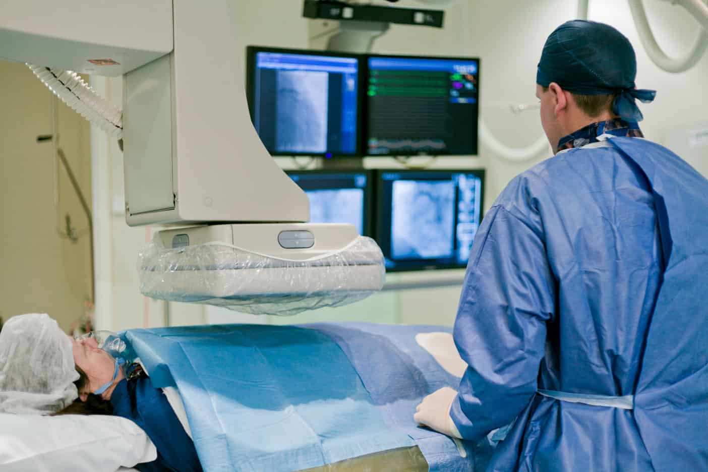 Interventional Radiology (IR) involves the use of advanced imaging technology and small medical instruments to diagnose and treat conditions inside the body. Many treatments that would have once required major surgery can now be done with interventional radiology procedures, which only require a small incision through the skin. Since these procedures are less invasive than surgery, they minimize the likelihood of infection and reduce recovery time.
Testing and Treatment for the Following Conditions:
- Abdominal Aortic Aneurysm
- Carotid Artery Disease
- Deep Venous Thrombosis
- Interventional Oncology
- Kidney Disease
- Liver Disease
- Peripheral Artery Disease
- Uterine Fibroids
- Thoracic Aortic Aneurysm
- Vascular Malformation
- Varicose Veins
- Vertebral Fracture
Interventional Radiology Procedures at KRMC:
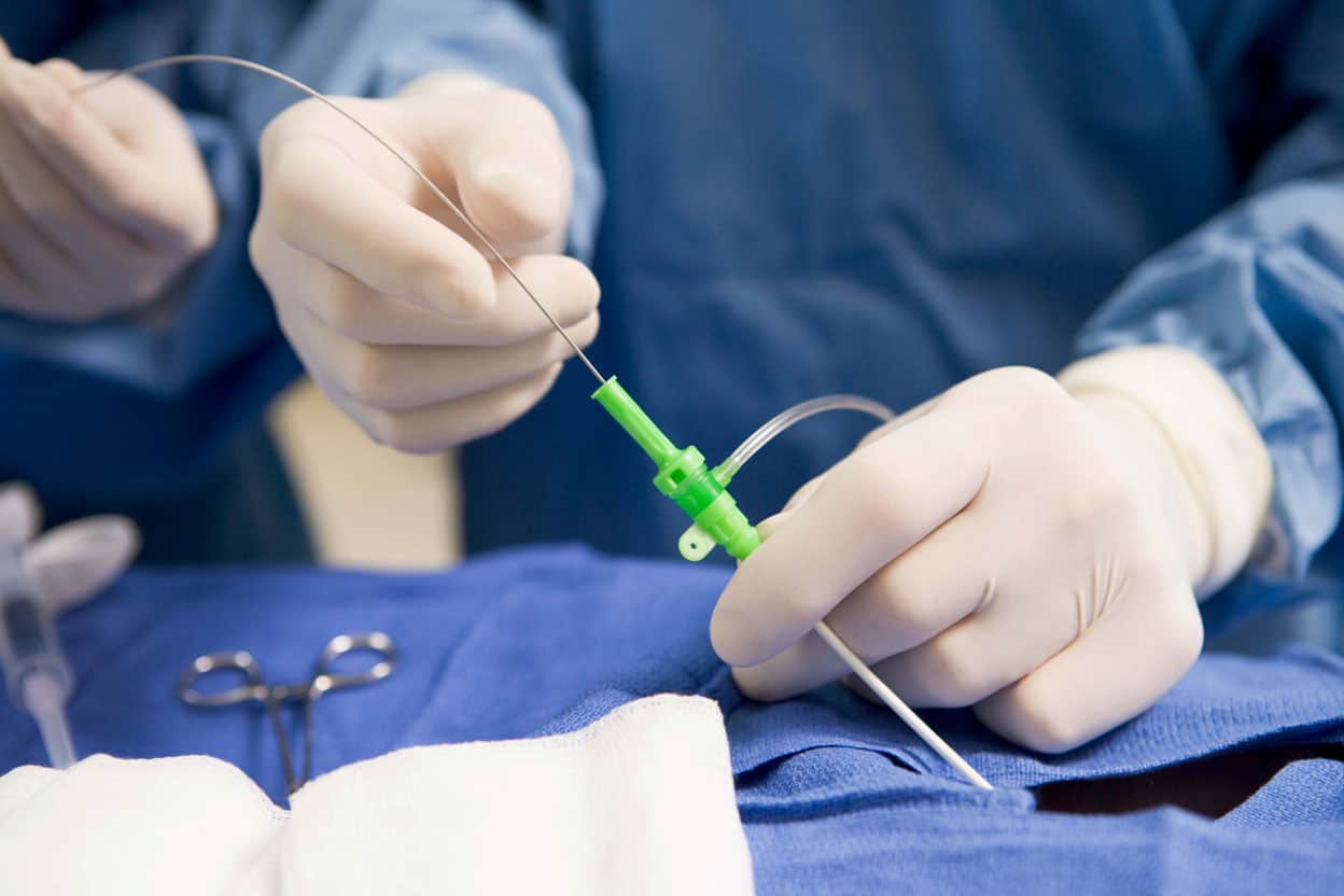 Atherectomy/Angioplasty Using X-ray guidance, an interventional radiologist will insert tiny tools through the skin, into the arteries, to treat a blocked or narrowed vessel. This procedure can be done for arteries in nearly every part of the body.
- Transjugular intrahepatic portosystemic shunt (TIPS)Using special tools, an interventional radiologist will place a shunt to create a pathway for blood to pass properly through a diseased liver. This procedure treats various conditions associated with liver disease including ascites and gastrointestinal bleeding.
- Aortic aneurysm repair (abdominal/thoracic) Endovascular aneurysm repair is a minimally invasive alternative to major open surgery for the repair of aortic aneurysms that result in reduced recovery times and potentially improved survival rates. Endovascular aneurysm repair is performed using a stent graft. The stent is placed within the aneurysm to provide a permanent, alternative channel for blood flow and preventing the walls of the aneurysm from rupturing. The stent graft is inserted into the aneurysm through small incisions in the groin without surgically opening or removing part of the aorta, thereby offering an alternative treatment choice to open surgery.
- Embolization An interventional radiologist can stop bleeding from damage to arteries by delivering gel, foam, and/or coils to the area using a catheter inserted through the skin. This procedure can also stop the flow of blood to other areas like an aneurysm or a fibroid tumor in the uterus. Additionally, chemotherapy drugs can be applied directly to a malignant tumor in a variation of this procedure called chemoembolization.
- Biopsy Using image guidance, an interventional radiologist can take a sample of tissue from the bones, bone marrow, liver, lung, lymph node, kidney, thyroid, and other areas of the body to evaluate and diagnose a mass or lesion.
- Port catheter An interventional radiologist will implant a port under the skin for long term access to the vein for patients who need chemotherapy and other repeated procedures.
- Central venous access The interventional radiologist will insert a tube through the skin and into the blood vessels for easier blood draws and delivery of medication.
- Vertebroplasty An interventional radiologist will use special tools to stabilize fractured vertebral bodies using bone cement.
- Drainage tubes An interventional radiologist will insert a catheter tube to drain excess fluid if the body is unable to drain the fluid itself. This is common in a few areas of the body. In the chest, the tube creates a pathway to remove air and/or fluid from the lung area. In the kidneys, nephrostomy tubes allow for internal or external drainage. In the bladder, suprapubic catheter provides an external drainage system.
- Arteriovenous (AV) fistulogram For patients on dialysis, an access is positioned under the skin to create an entryway to the bloodstream. An interventional radiologist can look for any narrowing or blockage in the dialysis access by inserting a catheter and injecting contrast dye for imaging. If necessary, the access can be repaired using special tools.