Offering the Latest Advances in Medical Imaging Technology and Expertise
Advances in modern imaging technologies have saved countless lives by detecting serious disease at earlier, more survivable stages. Kingman Regional Medical Center (KRMC) is proud to be the region’s leader in diagnostic imaging services. With the latest technology combined with the skilled expertise of our radiology team, patients at KRMC have access to some of modern medicine’s most-advanced imaging tests for diagnosing and treating illness and injury.
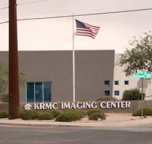
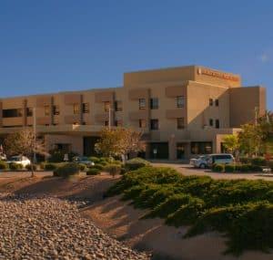
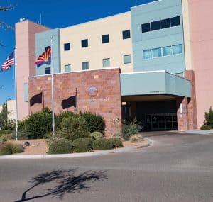
 Depending on the type of imaging test ordered by your doctor, we provide imaging services at the main hospital campus or at the KRMC Imaging Center. Our imaging services scheduler will inform you of the location when confirming your appointment.
Exceptional Radiology Expertise
Accurate imaging results require the expertise of doctors (called radiologists) who specialize in interpreting medical images for diagnosing and treating illness or injury. KRMC’s radiology team includes seven highly skilled radiologists who received their medical training at Mayo Clinic in Rochester, Minnesota. These doctors are skilled in the latest advances in medical imaging, including methods and techniques that reduce radiation exposure and improve patient safety. With their expertise and the latest imaging technologies, patients at KRMC have access to some of modern-medicine’s most advanced medical imaging services.
Imaging Capabilities at KRMC
Breast MRI Breast magnetic resonance imaging (MRI) is a test used to detect breast cancer and other abnormalities in the breast. A breast MRI captures multiple images of your breast. The images are combined, using a computer, to generate detailed pictures. In certain situations, such as for women with high risk of breast cancer, breast MRI may be used with mammograms as a screening tool for detecting breast cancer.
CT Scan A computerized tomography (CT) scan produces exceptionally clear three-dimensional images of structures inside your body. It is used to diagnose disease or injury or to plan medical or surgical treatment. It is particularly well-suited to quickly examine people who may have internal injuries from car accidents or other types of trauma. CT technology combines a series of X-ray images taken from different angles and uses computer processing to create cross-sectional images, or slices, of the bones, blood vessels and soft tissues inside your body.
DEXA Scan (Bone Density Test). A DEXA scan can determine if you have osteoporosis — a disease that causes bones to become more fragile and more likely to break. The test uses X-rays to measure how many grams of calcium and other bone minerals are packed into a segment of bone. You lie on a soft table. The scanner passes over your lower spine and hip. In most cases, you do not need to undress.
Image-guided needle biopsy An image-guided needle biopsy is a procedure that uses x-ray or ultrasound to precisely guide a special needle to a tumor or lesion in your body. The needle removes a small amount of tissue for laboratory testing.
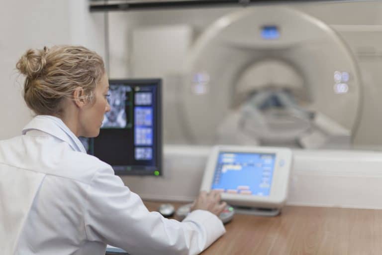
Magnetic Resonance Imaging (MRI). MRI is used to visualize soft tissue structures in the body. Unlike CT, it uses no ionizing radiation, but rather magnets and radio waves to create images. KRMC has two MRI systems, one of which is a powerful 3T MRI — one of only a few in Arizona, which produces a much stronger magnetic field than conventional MRI systems. It is highly useful in neurological (brain), musculoskeletal (body), cardiovascular (blood vessels and heart), and cancer imaging.
Mammography (3D Breast Tomosynthesis). A mammogram is an X-ray image of your breast used to screen for breast cancer. Mammograms play a key role in early breast cancer detection and help decrease breast cancer deaths. At KRMC, all mammograms are provided using advanced 3D mammography technology (called 3D breast tomosynthesis), which produces exceptionally clear three-dimensional images that enable doctors to see details in breast tissue in a way never before possible. Clinical studies show that 3D mammography significantly improves detection of early stage breast cancer, especially in women with dense breast tissue.
KRMC currently offers free 3D screening mammograms to women who reside in Mohave County through our Catch It Early program.
PET/CT scan KRMC’s Positron Emission Tomography/Computed Tomography (PET/CT) scanner is state-of-the-art technology that merges PET and CT together for showing how your tissues and organs are functioning. A PET/CT scan uses a radioactive drug (tracer) to show this activity. The tracer collects in areas of your body that have higher levels of chemical activity, which often correspond to areas of disease. On a PET/CT scan, these areas show up as bright spots.
PSMA PET scan This PET scan involves the use of a specialized radioactive tracer to detect prostate specific membrane antigen (PSMA) — a protein on the surface of prostate cancer cells. This allows doctors to determine the precise location of prostate cancer lesions, so they can create targeted treatment plans.
Ultrasound Ultrasound (also called sonography) is an imaging method that uses high-frequency sound waves to produce images of structures within your body. The images can provide valuable information for diagnosing and treating a variety of diseases and conditions.
X-ray An X-ray is a quick, painless test that produces images of structures inside your body — particularly your bones. X-ray beams pass through your body and they are absorbed in different amounts depending on the density of the material they pass through. Dense materials, such as bone and metal, show up as white on X-rays. The air in your lungs shows up as black. Fat and muscle appear as shades of gray. For some types of X-ray tests, a contrast medium — such as iodine or barium — is introduced into your body to provide greater detail on the images.